Mechanics and Synergistic Signaling of Fibronectin, Integrins, and TGF-β Isoforms

Review Open Access

Mechanics and Synergistic Signaling of Fibronectin, Integrins, and TGF-β Isoforms

Author Information
1
The Henry M. Jackson Foundation for the Advancement of Military Medicine, 6720A Rockledge Drive, Bethesda, MD 20817, USA
2
Department of Biomedical Engineering, Virginia Commonwealth University, 401 W. Main St., Richmond, VA 23284, USA
*
Authors to whom correspondence should be addressed.
Views:72
Downloads:11
Fibrosis 2025, 3 (1), 10003;  https://doi.org/10.70322/fibrosis.2025.10003

Received: 06 December 2024 Accepted: 27 January 2025 Published: 08 February 2025

Creative Commons

© 2025 The authors. This is an open access article under the Creative Commons Attribution 4.0 International License (https://creativecommons.org/licenses/by/4.0/).

ABSTRACT: Fibrotic diseases such as pulmonary fibrosis, hepatic fibrosis, chronic kidney disease, and cancer are marked by an excess accumulation of extracellular matrix (ECM). This process involves the assembly of the ECM protein fibronectin (FN) into insoluble fibrils. FN fibril assembly is highly linked with integrin signaling, TGF-β1 signaling, and cellular contractility. This linkage consists of four stages: (i) Integrin binding and contractile forces facilitate the assembly of FN into insoluble fibrils; (ii) assembled FN fibrils bind the large latent complex of TGF-β1; (iii) activation of TGF-β1 from the latent complex requires integrin binding and contractile forces; and (iv) active TGF-β1 increases contractility, integrin expression, and FN assembly. The significance of integrin signaling and TGF-β1 signaling in fibrotic diseases is well-appreciated, as numerous clinical trials targeting integrins or TGF-β1 have been reported. However, despite a clear effort to target integrins and TGF-β1 clinically, the vast majority of these trials have failed or have been terminated. These suggest a potentially incomplete understanding of the synergistic effects of these pathways. Here we present a review of both FN fibrillogenesis and TGF-β1 signaling, as well as current opinions of under-explored areas of crosstalk related to these pathways that may explain why these have not been successfully targeted in many disease states including fibrosis.
Keywords: Fibrosis; TGF-β; Fibronectin; Integrins mechanobiology

1. Introduction

1.1. TGF-β1 Activation and Signaling One of the most prominent inducers of fibrosis is the inflammatory cytokine Transforming Growth Factor-β1 (TGF-β1). In addition to its role as a modulator of immune system function (reviewed in [1]), TGF-β1 has been shown to play a significant role in a host of pathologies, including cancer tumorigenesis and metastasis, chronic kidney disease, idiopathic lung disease, cardiac disease, osteogenesis imperfecta, and Parkinson’s disease [2,3,4,5,6,7]. It is expressed in almost all tissues in the body (reviewed in [8]), and its expression is regulated by numerous signaling pathways (reviewed in [9]). TGF-β1 and its two related isoforms, TGF-β2 and TGF-β3, are secreted as latent complexes comprised of 3 components: a mature TGF-β ligand non-covalently bound to a latency-associated peptide (LAP), which is then bound to a latent TGF-β binding protein (LTBP). LTBP is able to bind to the extracellular matrix (ECM) protein fibronectin (FN) through cryptic growth factor binding sites on tensed FN, acting as a reservoir for latent TGF-β1 [10,11,12,13,14,15,16,17]. Active TGF-β1 can be released from the latent complex via decreases in pH, proteolysis by extracellular enzymes such as matrix metalloproteinases, or traction force-mediated opening of the LAP through αv integrins, most notably integrins αvβ1/3/6/8 [12,17,18,19]. These integrins are often clustered into large enzymatic complexes known as focal adhesions that coordinate extracellular attachments with the intracellular cytoskeleton (Figure 1). It is through these focal adhesions that intracellular, acto-myosin based traction forces are transduced to manipulate the ECM, regulate cell size, and coordinate cellular motility [12,14,20,21,22].
Figure 1
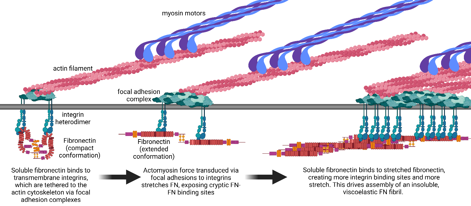
. Assembly of Fibronectin into Fibrils. Fibronectin is assembled into fibrils via an integrin-dependent mechanism. Integrins transmit actomyosin forces to soluble FN, which stretches the FN to reveal cryptic FN-FN binding sites. Binding of soluble FN to the stretched FN creates new integrin binding sites that facilitates the assembly of an insoluble FN fibril. Created with Biorender.com.
Active TGF-β ligands signal through a complex of cell surface receptors, comprising type I (TβRI), type II (TβRII), and type III (TβRIII) serine/threonine kinase receptors. During TGF-β signal transduction, a TGF-β ligand first binds to either a TβRII dimer or a pre-formed complex of a TβRI dimer and an RII dimer on the extracellular cell surface [23,24,25]. Ligand-bound TβRII then initiates recruitment of TβRI to form a heterotetramer complex. Constitutively active TβRII then phosphorylates the catalytic domain of TβRI on the cytoplasmic side, initiating downstream signal transduction [23,24]. TβRIII, also known as beta-glycan, can enhance the binding of TGF-β ligands to TβRII and is capable of complexing TβRI & II receptors together, serving as a co-receptor to increase TGF-β signaling in certain cell types [26,27,28]. While TGF-β2 has been implicated in fibrotic processes and TGF-β3 has been primarily identified as playing a role in physiological processes such as embryonic development, the TGF-β1 isoform has been characterized in the greatest detail in disease states [29]. TGF-β1 demonstrates the highest systemic abundance in the human body, with increases in expression associated with poor breast cancer prognosis and metastasis to bone, lymph, and pleural tissues [30,31,32]. It has also been implicated in Parkinson’s disease [3], fibrotic diseases [33], and osteogenesis imperfecta [7]. Each TGF-β isoform exhibits differences in ligand activation and receptor binding. TGF-β2 induces signals with 100–1000-fold less potency than TGF-β1 and TGF-β3 in cells that lack the noncatalytic TGF-βRIII (beta-glycan) co-receptor [23] and requires a preformed complex of TGF-βRI & TGF-βRII for sufficient ligand-receptor complexing [25,27]. TGF-β2 also lacks the typical integrin adhesion motif, RGD, and instead is only able to undergo integrin-mediated activation via an alternative recognition motif that is only recognized by integrin αvβ6 [34,35]. TGF-β3 has been shown to have a slightly greater affinity for TGF-βRII [23] than TGF-β1. TGF-β1 and 2 are known to promote scar formation during wound healing, while TGF-β3 promotes scar-less wound healing, indicating potentially divergent downstream signaling from TGF-β3 [36,37]. TGF-β signaling induces multiple downstream signaling events. In canonical TGF-β1 signaling, SMAD proteins are phosphorylated by the cytoplasmic domain of TβRI. SMAD2 and SMAD3 are the primary phosphorylation targets; these complex with SMAD4 and translocate to the nucleus to regulate target gene expression [24,25,26,27]. Noncanonical signaling also relies on signaling downstream of TβRI activity, but instead activates the mediators of the Rho family, NF-ΚB, Ras, MAPK, and PI3K signaling pathways to direct cytoskeletal rearrangement, protein synthesis, proliferation, and apoptosis [38,39]. 1.2. Fibronectin Fibrillogenesis and Regulation A primary component of fibrotic tissue is fibronectin (FN) [40,41], which serves both structural and signaling roles in de novo tissue morphogenesis. FN fibrils serve as scaffolds for the assembly of a variety of other ECM components, including collagens and elastins, facilitating the formation and maturation of new tissue [42,43,44]. This is an important point in fibrosis research: studies have shown that cells lacking FN are unable to assemble Collagen I fibrils in vivo [45,46,47]. This would suggest that inhibition of FN should also inhibit Collagen I assembly and, as such, serve as a novel target for fibrotic treatment. Indeed, treatment with an FN inhibitor in a unilateral ureteral obstruction (UUO) mouse model of kidney fibrosis demonstrated a significant reduction in Collagen I, Collagen III, and fibronectin in UUO kidneys [48]. In addition to serving roles as a structural element and a scaffold for Collagen I, assembled FN fibrils are also capable of binding upwards of 40 growth factors, including platelet-derived growth factors (PDGFs), fibroblast growth factors (FGFs), and of particular interest, latent TGF-β1 [10,15,41,49], making it a crucial regulator of cellular signal transduction in fibrotic foci. As such, FN fibrils not only dictate the mechanical structure of the fibrotic ECM, but also play a key role in regulating signaling cues to resident cells. FN is secreted as a soluble dimer that binds primarily to cell-surface integrin receptors, most notably integrins α5β1 and αvβ3. These integrins transmit actomyosin contractile forces to the FN molecule, which stretches FN into an open conformation that facilitates FN-FN binding [42,43]. This cycle of stretching to expose new binding sites and subsequent incorporation of soluble FN leads to the assembly of insoluble fibrils in a process known as fibrillogenesis [42,43,50] (Figure 1).The polymerization of FN into insoluble fibrillar networks serves as a scaffold for cellular adhesion, migration, and signaling, allowing for the subsequent association of other ECM constituents like collagens and elastins [43,49,51]. Further processing of FN fibrils includes both cleavage by proteolytic enzymes, which results in soluble fragments that exert their own cellular signaling function [52,53] and cross-linking by matricellular enzymes to reinforce attachments to both other FN fibrils and other ECM components. Both cleavage and cross-linking of FN fibrils dictate the bulk mechanical properties of the local ECM (reviewed in [43]). 1.3. Role of Integrins in FN Fibrillogenesis and TGF-β1 Activation A common thread between TGF-β1 activation and FN fibrillogenesis is that both require binding to transmembrane integrins and subsequent transmission of actomyosin-based contractile forces. For FN fibrillogenesis, both α5β1 and αvβ3 integrins bind to the RGD sequence on the 10th Type III domain of FN [54]. Transmission of actomyosin forces via these integrins stretches FN from a compact conformation to expose cryptic FN-FN binding sites that facilitate FN fibril formation [55]. While both α5β1 and αvβ3 integrins bind to FN, fibrillogenesis is primarily driven via α5β1 integrins [43]. For TGF-β1 activation, αv-containing integrins bind to the RGD sequence on the latency-associated peptide (LAP) of TGF-β1. Transmission of actomyosin forces via these integrins stretches LAP to release the active TGF-β1 ligand, allowing it to bind to TGF-β receptors and facilitate downstream signaling [56]. 1.4. Targeting Integrins and TGF-β1 Therapeutically The clinical significance of FN fibrillogenesis, TGF-β1 signaling, and integrin signaling in fibrotic diseases and cancer is evident by the vast number of clinical trials that have attempted to target these pathways [2,3,4,57,58,59,60,61,62]. Over 90 integrin-based therapeutics are under investigation, but only seven have been successfully marketed [58]. Targeting integrins has been proposed as a therapeutic approach to cancer [59,63] including glioblastoma [64], inflammatory bowel disease [57], cardiovascular disease [60], dry eye disease [60], idiopathic pulmonary fibrosis [62], and scleroderma fibrosis [65], amongst others. Similarly, targeting TGF-β has been a therapeutic target for a host of diseases, including cancer [2,4,5], Parkinson’s disease [3], and osteogenesis imperfecta [7]. 1.5. Underexplored Aspects of TGF-β-Integrin-FN Signaling The substantial number of clinical trials that have targeted either integrins or TGF-β1 indicate the significant role and pervasiveness of these pathways in fibrotic diseases; however, the large number of failures of these clinical trials [59,63,66] suggests an incomplete understanding of their complex signaling and crosstalk. Where are the shortcomings in understanding these signaling pathways? Here, we discuss three areas that may lie at the heart of this issue: (i) divergent regulation of TGF-β isoform expression in response to exogenous TGF-β1 depending on cell type; (ii) divergent FN expression and assembly responses to exogenous TGF-β1 depending on cell type and matrix microenvironment; and (iii) downstream integrin signaling in response to TGFβ/LAP binding to integrins.   1.6. Divergent Regulation of TGF-β Isoforms Exposure to exogenous TGF-β1 often leads to upregulation of endogenous expression of TGF-β isoforms, which has been hypothesized as an autocrine positive feedback loop that drives sustained TGF-β1 signaling and FN fibrillogenesis [67,68,69]. However, this response appears to be cell-type specific. Several studies have shown an upregulation of autocrine TGF-β1 in MDCK renal epithelial cells, MCF10 human mammary epithelial cells, and HK-2 human renal proximal tubular epithelial cells during TGF-β1-induced EMT [68,70,71], while others have shown no significant increase in autocrine TGF-β1 in MCF10A human mammary epithelial cells in response to pathological levels of exogenous TGF-β1 [69]. A possible theory to explain this discrepancy may be that TGF-β1 activity may predominantly be upregulated by increased accessibility of previously deposited latent TGF-β1 embedded in the ECM, which becomes increasingly accessible via upregulation of actomyosin contractility and matrix remodeling after initial TGF-β stimulation. Other studies have shown an upregulation of endogenous TGF-β2 in response to TGF-β1 in both MCF10A human mammary epithelial cells [69] and HCC 1954 human breast cancer cells [18], while other studies have shown a downregulation of TGF-β2 in response to TGF-β1 in MDA-MB-231 triple-negative breast cancer cells [69]. Interestingly, Barney et al. demonstrated that upregulation of TGF-β2 and FN correlated with cell survival in an in vitro model of tumor dormancy [18], while other studies indicated that TGF-β2 is elevated to significant levels in TNBC [72,73]. Divergent regulation of TGF-β isoforms may be regulated through several mechanisms. One avenue may be via posttranscriptional regulation of TGF-β translation. Liu et al. have proposed a mechanism of competitive endogenous FN mRNA expression, in which FN is transcribed solely as a mechanism to competitively bind miRNAs that inhibit TGF-β expression, thus upregulating TGF-β expression [67]. Another avenue for divergent responses to exogenous TGF-β1 is altered regulation of TGF-β receptors. As with most receptor-ligand pairs, TGF-β signaling is regulated by the internalization of receptors on the surface (reviewed in [74]). Specific regulation of receptors outside of clathrin-mediated mechanisms may play a key role in regulating divergent responses to TGF-β1. For example, β-arrestin2, which typically binds to 7-Transmembrane (7TM) receptors to drive internalization and inactivation of G-Protein Coupled Receptors, also mediates endocytosis of TβRIII through a similar mechanism to downregulate TGF-β1 signaling [75]. Collagen-α2β1 interactions downregulate TβR expression in a FAK-dependent mechanism in osteoblasts [76]; this further points to cross-talk between integrins and TGF-β signaling. The microRNA miR-150 drives expression of TGF-βRII in intra-epithelial lymphocytes to increase responsiveness to TGF-β1 [77]. 1.7. Divergent FN Expression and Assembly Previous studies have shown that mechanical activation of TGF-β1 requires tethering of the large latency complex to FN fibrils [10]. This is presumably due to the need for a resistive force on the LLC to facilitate mechanically induced opening of the LAP-TGF-β complex. Another avenue for divergent responses to exogenous TGF-β1 is distinct regulation of FN fibrillogenesis; inhibiting FN fibrillogenesis with a small bacterial wall peptide has been shown to inhibit TGF-β1 signaling [
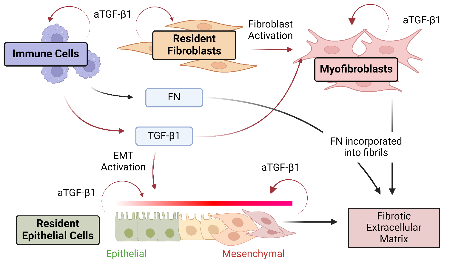
10]. The complexity of this aspect of FN-TGF-β1 crosstalk is amplified by the fact that FN fibrillogenesis requires both the secretion of soluble FN and the assembly of soluble FN into fibrils. This two-step process is not necessarily mediated by the same cell populations: one cell type could upregulate secretion of soluble FN while another could upregulate FN fibril assembly (and a third could upregulate expression of TGF-β1) (Figure 2).
Figure 2. Divergent sources of soluble FN, FN fibrillogenesis, and TGF-β1 expression in tissue microenvironments. A critical aspect of the complexity of FN/Integrin/TGF-β1 crosstalk is the potential for multiple cell types/cell phenotypes to contribute to the process. Multiple cell types, including immune cells, resident fibroblasts, and resident epithelial cells, respond to exogenous TGF-β1 by upregulating expression of soluble FN and endogenous, autocrine TGF-β1 (aTGF-β1). Transformed cells, including activated myofibroblasts and epithelial cells that have undergone Epithelial-Mesenchymal Transition (EMT), will assemble FN into fibrils. These fibrils can bind soluble TGF-β1, which can then be activated via integrin-transmitted contractile forces. Figure created with BioRender.com.
Assembly of FN fibrils seems to be more tightly regulated than expression of soluble FN, as there are many cell types and microenvironmental variables that drive increased expression of soluble FN without increased FN fibrillogenesis. Increased expression and secretion of soluble FN has been observed in MDA-MB-231 breast cancer cells [69], astrocytes exposed to an in vitro stroke model [78], hepatic stellate cells in a fibrogenesis animal model [79], HK-2 human renal proximal tubular epithelial cells in response to TGF-β1 [71], human lung epithelial cells in response to TGF-β1 [80], and CD14+ monocytes in systemic sclerosis [81]. These increases in soluble FN expression do not necessarily correlate with increased FN fibrillogenesis: for example, despite significant increases in FN secretion in response to TGF-β1, MDA-MB-231 cells do not readily assemble FN fibrils in 2D cultures [69], but do assemble fibrils in 3D assays [82]. Non-malignant MCF10A breast epithelial cells upregulate both FN secretion and assembly [10] in 2D. Hypoxia upregulates FN secretion in HK2 human kidney cells and A549 human lung adenocarcionma cells, but does not upregulate FN fibrillogenesis [83]. FN assembly is increased in mouse and human embryonic fibroblasts (3T3 and WI-38) in response to TGF-β1 [84,85]. FN assembly is also highly influenced by the abundance of certain ECM proteins in the matrix; culturing cells on laminin-coated surfaces increases FN fibrillogenesis in both MCF10A human epithelial cells [69] and NRK-49F rat renal fibroblasts (unpublished). There are several potential mechanisms that could explain how cells facilitate FN fibrillogenesis independent of FN expression and secretion. TGF-β1 induces an increase in surface transglutaminase in mouse and human embryonic fibroblasts (3T3 and WI-38), which co-localizes with α5β1 integrins and may play a role in localizing soluble FN to integrins on the cell surface [84]. TβRIII promotes epithelial cell attachment to FN by trafficking α5β1 integrins to nascent adhesions [86]; this effect is independent of exogenous β1. Inhibiting α4 integrin disrupts the binding of the EDA domain of FN to integrins; this results in reduced contractility and reduced FN fibrillogenesis [87]. Note that the EDA domain is not present in all isoforms of FN; it is an alternatively spliced domain that is not present in systemic FN expressed in hepatocytes (“plasma FN”) but is often present in locally expressed FN (“cellular FN”). As such, divergent expression of transglutaminase, TβRIII, α4 integrin, and/or EDA-containing isoforms of FN could play a role in divergent FN fibrillogenesis responses to exogenous TGF-β1. 1.8. Downstream Integrin-LAP Signaling 
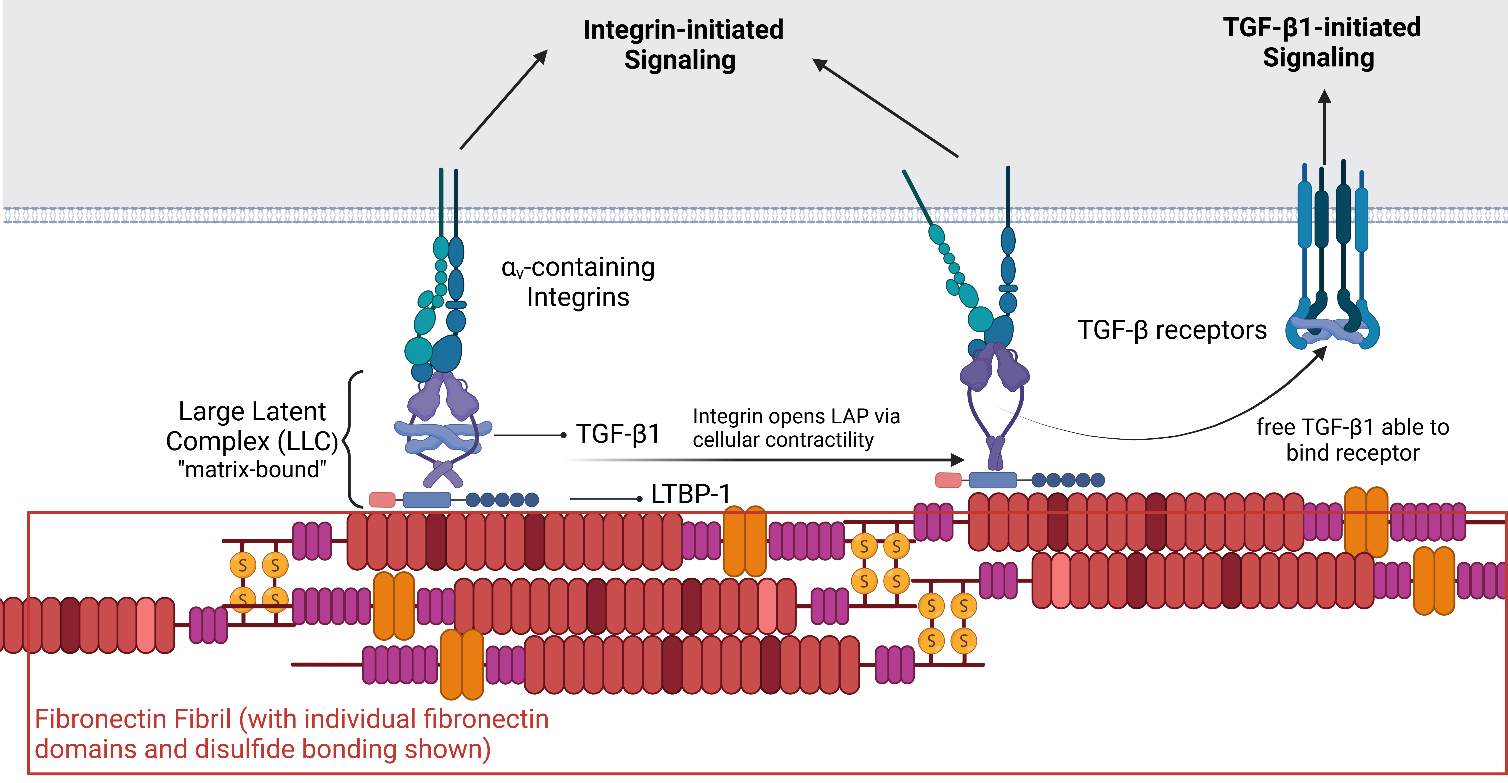
Another potentially important and under-explored avenue of the TGF-β1/FN/Integrin axis is the interaction between the TGF-β1 LAP protein and integrins (Figure 3). While the importance of this binding is well appreciated and has been explicitly targeted in clinical trials, the focus has been on the role of integrin-LAP binding in facilitating mechanical forces that physically release the active TGF-β ligand. However, LAP-integrin binding may also serve as a traditional receptor-ligand binding event that triggers signaling events downstream of integrin ligation. Results of several studies suggest that this mechanism may play a key role. Studies from our group found that knocking out TGF-β1 resulted in impaired FN fibrillogenesis in MCF10A human mammary epithelial cells, which was not recovered by the addition of soluble TGF-β1. However, addition of the empty LAP protein to TGF-β1 knock out cells did partially recover FN fibrillogenesis [62]. RGD-binding integrins have the greatest affinity for LAP in its empty conformation (that is, with no TGF-β ligand contained within it), highlighting a potential role in modulating cellular signaling. Prior studies have demonstrated that empty LAP facilitates keratinocyte adhesion [88] and modulates monocyte chemotaxis and inflammation response [89].
Figure 3. Crosstalk between integrin signaling, TGF-β1 signaling, and Fibronectin. Integrins bind to the Large Latent Complex (LLC) that contains the active TGF-β ligand; the LLC can either be soluble or bound to FN fibrils. Bound LLC is stretched via actomyosin forces that open the LLC and release the active TGF-β ligand. This ligand can then bind TGF-β receptors. Integrins bound to soluble LLC, matrix-bound LLC, and/or opened LLC may drive intracellular signaling that augments signaling downstream of the TGF-β1 ligand. Created with BioRender.com.

2. Conclusions

Crosstalk between integrins, FN, and TGF-β1 is critical for both physiological processes, including wound healing, and enbryogenesis and pathological scenarios, including fibrotic diseases and cancer. While numerous therapeutic agents have been developed to target these, most have failed. The complex interactions described here lead to an unclear path to specifically target the role of these signaling moieties in disease states without disrupting their role in healthy environments. We believe that the answer to this conundrum lies in three areas: (i) identify mechanisms that specifically regulate TGF-β1 responses, including modulation of receptors; (ii) identify mechanisms that regulate FN fibrillogenesis, and specifically target this in pathological environments (as has been attempted by [48]), and (iii) identify signaling events downstream of LAP-integrin binding that may be specifically critical to long-term pathological fibrotic signaling, given the absence of LAP in immune cell-derived TGF-β1.

Author Contributions

Writing—Original Draft Preparation, M.M.S., C.A.L.; Writing—Review and Editing: M.M.S., C.A.L.; Project Administration, C.A.L.; Funding Acquisition, C.A.L.

Ethics Statement

Not applicable.

Informed Consent Statement

Not applicable.

Data Availability Statement

Not applicable.

Funding

This research was supported by the National Science Foundation via Award CBET-2302580 (M.M.S., C.A.L.).

Declaration of Competing Interest

The authors declare that they have no known competing financial interests or personal relationships that could have appeared to influence the work reported in this paper.

References

1.
Massague J, Sheppard D. TGF-β signaling in health and disease. Cell 2023, 186, 4007–4037. [Google Scholar]
2.
Akhurst RJ. Targeting TGF-β signaling for therapeutic gain. Cold Spr. Harb. Perspect. Biol. 2017, 9, a022301. [Google Scholar]
3.
Karampetsou M, Vekrellis K, Melachroinou K. The promise of the TGF-β superfamily as a therapeutic target for Parkinson’s disease. Neur. Dis. 2022, 171, 105805. [Google Scholar]
4.
Hawinkels LJ, Dijke PT. Exploring anti-TGF-β therapies in cancer and fibrosis. Growth Fact. 2011, 29, 140–152. [Google Scholar]
5.
Smith AL, Robin TP, Ford HL. Molecular pathways: Targeting the TGF-β pathway for cancer therapy. Clin. Cancer Res. 2012, 18, 4514–4521. [Google Scholar]
6.
Tie Y, Tang F, Peng D, Zhang Y, Shi H. TGF-beta signal transduction: biology, function and therapy for diseases. Mol. Biomed. 2022, 3, 45. [Google Scholar]
7.
Song IW, Nagamani SC, Nguyen D, Grafe I, Sutton VR, Gannon FH, et al. Targeting TGF-β for treatment of osteogenesis imperfecta. J. Clin. Investig. 2022, 7, 132. 2022, 7, 132. [Google Scholar]
8.
Wang CZ, Zhang ZQ, Zhang Y, Zheng LF, Liu Y, Yan AT, et al. Comprehensive characterization of TGFB1 across hematological malignancies. Sci. Rep. 2023, 13, 19107. [Google Scholar]
9.
Hariyanto NI, Yo EC, Wanandi SI. Regulation and Signaling of TGF-β Autoinduction. Int. J. Mol. Cell. Med. 2022, 10, 234. [Google Scholar]
10.
Griggs LA, Hassan NT, Malik RS, Griffin BP, Martinez BA, Elmore LW, et al. Fibronectin fibrils regulate TGF-β1-induced Epithelial- Mesenchymal Transition. Matrix Biol. 2017, 60, 157–175. [Google Scholar]
11.
Wang JP, Hielscher A. Fibronectin: How Its Aberrant Expression in Tumors May Improve Therapeutic Targeting. J. Cancer 2017, 8, 674–682. [Google Scholar]
12.
Hinz B. The extracellular matrix and transforming growth factor-β1: Tale of a strained relationship. Matrix Biol. 2015, 47, 54–65. [Google Scholar]
13.
Zhu J, Clark RA. Fibronectin at select sites binds multiple growth factors and enhances their activity: Expansion of the collaborative ECM-GF paradigm. J. Investig. Dermatol. 2014, 134, 895–901. [Google Scholar]
14.
Hargadon K. Dysregulation of TGFβ1 Activity in Cancer and Its Influence on the Quality of Anti-Tumor Immunity. J. Clin. Med. 2016, 5, 76. [Google Scholar]
15.
Martino MM, Hubbell JA. The 12th–14th type III repeats of fibronectin function as a highly promiscuous growth factor-binding domain. FASEB J. 2010, 24, 4711–4721. [Google Scholar]
16.
Zhong X, Arnolds O, Krenczyk O, Gajewski J, Putz S, Herrmann C, et al. The Structure in Solution of Fibronectin Type III Domain 14 Reveals Its Synergistic Heparin Binding Site. Biochemistry 2018, 57, 6045–6049. [Google Scholar]
17.
Miyamoto S, Teramoto H, Gutkind JS, Yamada KM. Integrins can collaborate with growth factors for phosphorylation of receptor tyrosine kinases and MAP kinase activation: roles of integrin aggregation and occupancy of receptors. J. Cell Biol. 1996, 135, 1633. [Google Scholar]
18.
Barney LE, Hall CL, Schwartz AD, Parks AN, Sparages C, Galarza S, et al. Tumor cell–organized fibronectin maintenance of a dormant breast cancer population. Sci. Adv. 2020, 6, eaaz4157. [Google Scholar]
19.
Kobayashi T, Kim H, Liu X, Sugiura H, Kohyama T, Fang Q, et al. Matrix metalloproteinase-9 activates TGF-β and stimulates fibroblast contraction of collagen gels. Am. J. Phy. Lung Cell. Mol. Phy. 2014, 306, L1006–L1015. [Google Scholar]
20.
Santibanez JF, Obradovic H, Kukolj T, Krstic J. Transforming growth factor-β, matrix metalloproteinases, and urokinase-type plasminogen activator interaction in the cancer epithelial to mesenchymal transition. Develop. Dynam. 2018, 247, 382–395. [Google Scholar]
21.
Furler RL, Nixon DF, Brantner CA, Popratiloff A, Uittenbogaart CH. TGF-β sustains tumor progression through biochemical and mechanical signal transduction. Cancers 2018, 10, 199. [Google Scholar]
22.
Kim DH, Wirtz D. Predicting how cells spread and migrate: Focal adhesion size does matter. Cell Adhes. Migrat. 2013, 7, 293. [Google Scholar]
23.
Huang T, Schor SL, Hinck AP. Biological Activity Differences between TGF-β1 and TGF-β3 Correlate with Differences in the Rigidity and Arrange- ment of Their Component Monomers. Biochemistry 2014, 53, 5737. [Google Scholar]
24.
Vizán P, Miller DS, Gori I, Das D, Schmierer B, Hill CS. Controlling Long-Term Signaling: Receptor Dynamics Determine Attenuation and Re- fractory Behavior of the TGF-β Pathway. Sci. Signal. 2013, 6, 106. [Google Scholar]
25.
Rodriguez C, Chen F, Weinberg RA, Lodish HF. Cooperative Binding of Transforming Growth Factor (TGF)-β2 to the Types I and II TGF-β Receptors. J. Biol. Chem. 1995, 270, 15919–15922. [Google Scholar]
26.
Rys JP, DuFort CC, Monteiro DA, Baird MA, Oses-Prieto JA, Chand S, et al. Discrete spatial organi- zation of TGFβ receptors couples receptor multimer- ization and signaling to cellular tension. eLife 2015, 4, e09300. [Google Scholar]
27.
Wells RG, Yankelev H, Lin HY, Lodish HF. Biosynthesis of the type I and type II TGF-β receptors: implications for complex formation. J. Biol. Chem. 1997, 272, 11444–11451. [Google Scholar]
28.
Liu Z, Li M, Jiang Z, Wang X. A Comprehen- sive Immunologic Portrait of Triple-Negative Breast Cancer. Trans. Oncol. 2018, 11, 311–329. [Google Scholar]
29.
Morikawa M, Derynck R, Miyazono K. TGF- β and the TGF-β family: Context-dependent roles in cell and tissue physiology. Cold Spr. Harb. Perspect. Biol. 2016, 8, a021873. [Google Scholar]
30.
Mourskaia AA, Dong Z, Ng S, Banville M, Zwaagstra JC, O'Connor-McCourt MD, et al. Transforming growth factor-β1 is the predominant isoform required for breast cancer cell outgrowth in bone. Oncogene 2009, 28, 1005–1015. [Google Scholar]
31.
Park J, Schwarzbauer JE. Mammary epithelial cell interactions with fibronectin stimulate epithelial- mesenchymal transition. Oncogene 2014, 33, 1649–1657. [Google Scholar]
32.
Li CL, Yang D, Cao X, Wang F, Hong DY, Wang J, et al. Fibronectin induces epithelial-mesenchymal transition in human breast cancer MCF-7 cells via activation of calpain. Oncol. Lett. 2017, 13, 3889. [Google Scholar]
33.
Leask A, Abraham DJ. TGF-β signaling and the fibrotic response. FASEB J. 2004, 18, 816–827. [Google Scholar]
34.
Nishimura SL. Integrin-Mediated Transforming Growth Factor-β Activation, a Potential Therapeutic Target in Fibrogenic Disorders. Am. J. Pathol. 2009, 175, 1362. [Google Scholar]
35.
Le VQ, Zhao B, Ramesh S, Toohey C, DeCosta A, Mintseris J, et al. A specialized integrin-binding motif enables proTGF-β2 activation by integrin αVβ6 but not αVβ8. Proc. Nat. Acad. Sci. USA 2023, 6, 120. [Google Scholar]
36.
Ferguson MW, O'Kane S. Scar-free healing: From embryonic mechanism to adult therapeutic in- tervention. Philo. Trans. Royal Soc. Biol. Sci. 2004, 359, 839–850. [Google Scholar]
37.
Shah M, Foreman D, Ferguson M. Neutralisation of TGF-beta 1 and TGF-beta 2 or exogenous addition of TGF-beta 3 to cutaneous rat wounds reduces scarring. J. Cell Sci. 1995, 108, 985–1002. [Google Scholar]
38.
Alvarez MA, Freitas JP, Hussain SM, Glazer ES. TGF-β Inhibitors in Metastatic Pancreatic Ductal Adenocarcinoma. J. Gast. Cancer 2019, 50, 207–213. [Google Scholar]
39.
Finnson KW, Almadani Y, Philip A. Non- canonical (non-SMAD2/3) TGF-β signaling in fibro- sis: Mechanisms and targets. Semi. Cell Develop. Biol. 2020, 101, 115–122. [Google Scholar]
40.
Huang J, Zhang L, Wan D, Zhou L, Zheng S, Lin S, et al. Extracellular matrix and its therapeutic potential for cancer treatment. Signal Trans. Target. Ther. 2021, 6, 1–24. [Google Scholar]
41.
Yoneda A. Fibronectin Matrix Assembly and Its Significant Role in Cancer Progression and Treatment. Trends Glycosci. Glycotech. 2015, 27, 89–98. [Google Scholar]
42.
Schwarzbauer JE, DeSimone DW. Fibronectins, Their Fibrillogenesis, and In Vivo Functions. Cold Spr. Harbor Perspect. Biol. 2011, 3, 1–19. [Google Scholar]
43.
Dalton CJ, Lemmon CA. Fibronectin: Molecular Structure, Fibrillar Structure, and Mechanochemical Signaling. Cells 2021, 10, 2443. [Google Scholar]
44.
Libring S, Shinde A, Chanda MK, Nuru M, George H, Saleh AM, et al. The dy- namic relationship of breast cancer cells and fibroblasts in fibronectin accumulation at primary and metastatic tumor sites. Cancers 2020, 12, 1270. [Google Scholar]
45.
Sottile J, Shi F, Rublyevska I, Chiang HY, Lust J, Chandler J. Fibronectin-dependent collagen I deposition modulates the cell response to fibronectin. Am. J. Phy. Cell Phy. 2007, 293, C1934–C1946. [Google Scholar]
46.
Kadler KE, Hill A, Canty-Laird EG. Collagen fibrillogenesis: fibronectin, integrins, and minor collagens as organizers and nucleators. Curr. Opin. Cell Biol. 2008, 20, 495–501. [Google Scholar]
47.
Shi F, Harman J, Fujiwara K, Sottile J. Collagen I matrix turnover is regulated by fibronectin polymerization. Am. J. Phy. Cell Phy. 2010, 298, C1265–C1275. [Google Scholar]
48.
Tomasini-Johansson BR, Mosher DF. Microtiter assays for quantitation of assembly of plasma and cellular fibronectin. Meth. Cell Biol. 2018, 143, 157–170. [Google Scholar]
49.
Ohashi T, Erickson HP. Fibronectin aggregation and assembly: The unfolding of the second fibronectin type III domain. J. Biol. Chem. 2011, 286, 39188–39199. [Google Scholar]
50.
Weinberg SH, Mair DB, Lemmon CA. Mechanotransduction Dynamics at the Cell-Matrix Interface. Biophy. J. 2017, 112, 1962. [Google Scholar]
51.
Lemmon CA, Ohashi T, Erickson HP. Probing the folded state of fibronectin type III domains in stretched fibrils by measuring buried cysteine accessibility. J. Biol. Chem. 2011, 286, 26375–26382. [Google Scholar]
52.
Ruel N, Markova DZ, Adams SL, Scanzello C, Cs-Szabo G, Gerard D, et al. Fibronectin Fragments and the Cleaving Enzyme ADAM-8 in the Degenerative Human Intervertebral Disc. Spine 2014, 39, 1274–1279. [Google Scholar]
53.
Joshi R, Goihberg E, Ren W, Pilichowska M, Mathew P. Proteolytic fragments of fibronectin function as matrikines driving the chemotactic affinity of prostate cancer cells to human bone marrow mesenchy- mal stromal cells via the α5β1 integrin. Cell Adhes. Migrat. 2017, 11, 305–315. [Google Scholar]
54.
Ruoslahti E, Pierschbacher MD. Arg-Gly-Asp: A versatile cell recognition signal. Cell 1986, 44, 517–518. [Google Scholar]
55.
Zhong C, Chrzanowska-Wodnicka M, Brown J, Shaub A, Belkin AM, Burridge K.  Rho-mediated contractility exposes a cryptic site in fibronectin and induces fibronectin matrix assembly. Cell J. Biol. 1998, 141, 539–551. [Google Scholar]
56.
Wipff PJ, Hinz B. Integrins and the activation of latent transforming growth factor β1-An intimate relationship. Eur. J. Cell Biol. 2008, 87, 601–615. [Google Scholar]
57.
Solitano V, Parigi TL, Ragaini E, Danese S. Anti-integrin drugs in clinical trials for inflammatory bowel disease (IBD): insights into promising agents. Exp. Opi. Invest. Drugs 2021, 30, 1037–1046. [Google Scholar]
58.
Pang X, He X, Qiu Z, Zhang H, Xie R, Liu Z, et al. Targeting integrin pathways: mechanisms and advances in therapy. Signal Trans. Targ. Therapy 2023, 8, 1–42. [Google Scholar]
59.
Alday-Parejo B, Stupp R, Rüegg C. Are integrins still practicable targets for anti-cancer therapy? Cancers 2019, 11, 978. [Google Scholar]
60.
Slack RJ, Macdonald SJ, Roper JA, Jenkins RG, Hatley RJ. Emerging therapeutic opportunities for integrin inhibitors. Nat. Rev. Drug Dis. 2022, 21, 60–78. [Google Scholar]
61.
Ludwig BS, Kessler H, Kossatz S, Reuning U. Rgd-binding integrins revisited: How recently discov- ered functions and novel synthetic ligands (re-)shape an ever-evolving field. Cancers 2021, 13, 1711. [Google Scholar]
62.
Sime P, Jenkins G. Goldilocks and the Three Trials: Clinical Trials Targeting the αvβ6 Integrin in Idiopathic Pulmonary Fibrosis. Am. J. Respirat. Crit. Care Med. 2022, 206, 1062–1063. [Google Scholar]
63.
Bergonzini C, Kroese K, Zweemer AJ, Danen EH. Targeting Integrins for Cancer Therapy-Disappointments and Opportunities. Front. Cell Develop. Biol. 2022, 10, 863850. [Google Scholar]
64.
Malric L, Monferran S, Gilhodes J, Boyrie S, Dahan P, Skuli N, et al.  Interest of integrins targeting in glioblastoma according to tumor heterogeneity and cancer stem cell paradigm: An update. Oncotarget 2017, 8, 86947. [Google Scholar]
65.
Leask A, Fadl A, Naik A. A modest proposal: targeting αv integrin-mediated activation of la- tent TGFbeta as a novel therapeutic approach to treat scleroderma fibrosis. Exp. Opin. Invest. Drugs 2024, 33, 279–285. [Google Scholar]
66.
Zhang M, Zhang YY, Chen Y, Wang J, Wang Q, Lu H. TGF-β Signaling and Resistance to Cancer Therapy. Front. Cell Develop. Biol. 2021, 9, 786728. [Google Scholar]
67.
Liu Y, Xue M, Du S, Feng W, Zhang K, Zhang L, et al. Competitive endogenous RNA is an intrinsic component of EMT regulatory circuits and modulates EMT. Nat. Commun. 2019, 10, 1–12. [Google Scholar]
68.
Gregory PA, Bracken CP, Smith E, Bert AG, Wright JA, Roslan S, et al. An au- tocrine TGF-β/ZEB/miR-200 signaling network reg- ulates establishment and maintenance of epithelial- mesenchymal transition. Mol. Biol. Cell 2011, 22, 1686. [Google Scholar]
69.
Sofroniou MM, Lemmon CA. Differential reg- ulation of fibronectin expression and fibrillogenesis by autocrine TGF-β1 signaling in malignant and benign mammary epithelial cells. Inter. J. Biochem. Cell Biol. 2023, 165, 106478. [Google Scholar]
70.
Zhang J, Tian XJ, Zhang H, Teng Y, Li R, Bai F, et al. TGF-β-induced epithelial-to-mesenchymal transition proceeds through stepwise activation of multiple feedback loops. Sci. Signal. 2014, 7, ra91. [Google Scholar]
71.
Garmaa G, Manzeger A, Haghighi S, Kokeny G. HK-2 cell response to TGF-β highly depends on cell culture medium formulations. Histochem. Cell Biol. 2024, 161, 69–79. [Google Scholar]
72.
Xu X, Zhang L, He X, Zhang P, Sun C, Xu X, et al. TGF-beta plays a vital role in triple-negative breast cancer (TNBC) drug-resistance through regulating stemness, EMT and apoptosis. Biochem. Biophy. Res. Commun. 2018, 502, 160–165. [Google Scholar]
73.
Kim S, Lee J, Jeon M, Nam SJ, Lee JE. Elevated TGF-β1 and -β2 expression accelerates the epithelial to mesenchymal transition in triple-negative breast cancer cells. Cytokine 2015, 75, 151–158. [Google Scholar]
74.
Huang F, Chen YG. Regulation of TGF-β receptor activity. Cell Biosci. 2012, 2, 1–10. [Google Scholar]
75.
Chen W, Kirkbride KC, How T, Nelson CD, Mo J, Frederick JP, et al. β-arrestin 2 mediates endocytosis of type III TGF-β receptor and down-regulation of its signaling. Science 2003, 301, 1394–1397. [Google Scholar]
76.
Takeuchi Y, Suzawa M, Kikuchi T, Nishida E, Fujita T, Matsumoto T. Differentiation and trans- forming growth factor-β receptor downregulation by collagen-α2β1 integrin interaction is mediated by focal adhesion kinase and its downstream signals in murine osteoblastic cells. J. Biol. Chem. 1997, 272, 29309–29316. [Google Scholar]
77.
Seo SH, Jang MS, Kim DJ, Kim SM, Oh SC, Jung CR, et al. MicroRNA-150 controls differentiation of intraepithelial lymphocytes through TGF-β receptor II regulation. J. Allergy Clin. Immunol. 2018, 141, 1382–1394. [Google Scholar]
78.
Howe MD, Furr JW, Munshi Y, Roy-O’Reilly MA, Maniskas ME, Koellhoffer EC, et al. Transforming growth factor-β promotes basement membrane fibrosis, alters perivascular cerebrospinal fluid distribution, and worsens neurological recovery in the aged brain after stroke. GeroScience 2019, 41, 543–559. [Google Scholar]
79.
Liu CY, Lin HH, Tang MJ, Wang YK. Vimentin contributes to epithelial-mesenchymal transition cancer cell mechanics by mediating cytoskeletal organization and focal adhesion maturation. Oncotarget 2015, 6, 15966. [Google Scholar]
80.
Cheng WH, Kao SY, Chen CL, Yuliani FS, Lin LY, Lin CH, et al. Amphiregulin induces CCN2 and fibronectin expression by TGF-β through EGFR-dependent pathway in lung epithelial cells. Respirat. Res. 2022, 23, 381. [Google Scholar]
81.
Rudnik M, Hukara A, Kocherova I, Jordan S, Schniering J, Milleret V, et al. Elevated Fibronectin Levels in Profibrotic CD14+ Monocytes and CD14+ Macrophages in Systemic Sclerosis. Front. Immunol. 2021, 12, 642891. [Google Scholar]
82.
Park HJ, Helfman DM. Up-regulated fibronectin in 3D culture facilitates spreading of triple negative breast cancer cells on 2D through integrin β-5 and Src. Sci. Rep. 2019, 9, 1–14. [Google Scholar]
83.
Rana MK, Srivastava J, Yang M, Chen CS, Barber DL. Hypoxia increases the abundance but not the assembly of extracellular fibronectin during epithelial cell transdifferentiation. J. Cell Sci. 2015, 128, 1083–1089. [Google Scholar]
84.
Akimov SS, Belkin AM. Cell-surface transglu- taminase promotes fibronectin assembly via interaction with the gelatin-binding domain of fibronectin: A role in TGF-β-dependent matrix deposition. J. Cell Sci. 2001, 114, 2989–3000. [Google Scholar]
85.
Scott L, Mair D, Narang J, Feleke K, Lemmon CA. Fibronectin fibrillogenesis facilitates mechano-dependent cell spreading, force generation, and nuclear size in human embryonic fibroblasts. Integr. Biol. 2015, 7, 1454. [Google Scholar]
86.
Mythreye K, Knelson EH, Gatza CE, Gatza ML, Blobe GC. TβRIII/β-arrestin2 regulates integrin α5β1 trafficking, function, and localization in epithelial cells. Oncogene 2013, 32, 1416–1427. [Google Scholar]
87.
Shinde AV, Kelsh R, Peters JH, Sekiguchi K, Van De Water L, McKeown-Longo PJ. The α4β1 integrin and the EDA domain of fibronectin regulate a profibrotic phenotype in dermal fibroblasts. Matrix Biol. 2015, 41, 26–35. [Google Scholar]
88.
Thomas GJ, Hart IR, Speight PM, Marshall JF. Binding of TGF-β1 latency-associated peptide (LAP) to αvβ6 integrin modulates behaviour of squamous carcinoma cells. Brit. J. Cancer 2002, 87, 859–867. [Google Scholar]
89.
Ali NA, Gaughan AA, Orosz CG, Baran CP, McMaken S, Wang Y, et al. Latency Associated Peptide Has In Vitro and In Vivo Immune Effects Independent of TGF-β1. PLoS ONE 2008, 3, 1914. [Google Scholar]
TOP