Article Open Access
Received: 03 December 2024 Accepted: 13 February 2025 Published: 19 February 2025

© 2025 The authors. This is an open access article under the Creative Commons Attribution 4.0 International License (https://creativecommons.org/licenses/by/4.0/).
Nickel, iron, and cobalt nanoparticles have attracted increasing interest among researchers because they have ferro-magnetic properties at room temperature [1]. Recently, cobalt oxides (Co3O4) have received considerable attention in various fields due to their unique antiferromagnetic p-type semiconductor properties and possible applications in magnetic fluids, catalysis, biotechnology, magnetic resonance imaging (MRI), and data storage [2,3,4,5]. When reduced down to the nanometer scale, (Co3O4·NPs) were found to have interesting magnetic, optical, field emission, and electrochemical properties that are attractive in device applications [6,7].
Sonochemical synthesis offers a unique way to create known compounds without requiring very high temperatures, high pressures, and/or lengthy reaction durations. It is nearly a current technology for producing new materials [8]. The chemical reactions of molecules that are triggered by acoustic cavitation under ultrasonic radiation (20–10 MHz) are linked to the mechanism of sonochemistry [9,10]. The creation and expansion of bubbles in a liquid medium, followed by the implosive collapse of overgrown bubbles, are the phases of acoustic cavitation. High pressures and strong local temperatures are also present throughout this process [11,12]. Comparing the sonochemical approach to other methods, its benefits include ease of use, a quick reaction time, non-thermal processing, and the lack of extra cross-linking agents [13,14]. Thus, it contributes with the surfactant to produce nanoparticles with better properties and features.
As a surfactant, PEG is one of the polymers with major interest in this area because it is nontoxic, non-flammable and easy to handle. It has been reported that PEG with a uniform and ordered chain structure is easily adsorbed at the surface of metal oxide colloid. The addition of (PEG) to the reaction solution will alter the growth process’ kinetics, which is linked to nucleation’s quick expansion and the aggregation of nanoparticles [15]. Additionally, a number of researchers have documented the synthesis of noble metal nanoparticles using PEG, taking advantage of its non-irritating, non-toxic, and moisturizing characteristics [16,17].
PEG is a versatile polymer that has extensive applications in green chemistry, cosmetics, and pharmaceuticals [18,19]. Wide molecular weight ranges, flexibility, and biocompatibility are some of its main advantages. PEG stabilizes proteins, enhances medication administration, and facilitates chromatographic separations. As a solvent in environmentally friendly synthesis, it also serves as an emulsifier in cosmetics. PEGylation improves the stability of drugs, but it also presents problems with immunogenicity and molecular weight regulation. Its safety and uses are being optimized through ongoing study [20,21,22].
In this work, we present an entirely new approach involving less energy and low-cost coating material (polyethylene-glycol (PEG)) stabilized (Co3O4·NPs) assisted sonochemical method, its structural, spectroscopic analysis, morphological and optical properties were presented.
X-ray diffraction patterns of (Co3O4·NPs) are shown in Figure 1. It can be clearly identified that the production of cobalt oxide nanoparticles with polyethylene glycol as surfactant (Co3O4·NPs + PEG) has shown the most nanostructures corresponding to the cubic spinel nanocrystal phase (JCPDS No. 03-065-3103, crystal system: cubic, space group: Fd-3m). The main characteristic peaks are near to (2θ = 19.13°, 31.33°, 36.89°, 38.55°, 44.79°, 55.67°, 59.47°, 65.40°, and 77.34°) corresponding to the planes (111), (220), (311), (222), (400), (422), (511), (440) and (533). The considerable broadening of the diffraction peaks indicated the nanocrystal nature of the cobalt oxide (Co3O4) particles. The average crystallite size of the cobalt oxide nanoparticles (Co3O4·NPs) prepared with polyethylene glycol (PEG) surfactant was around 15 nm, which was determined from the X-ray diffraction analysis (XRD) pattern parameters according to the classical Debye–Scherrer Equation (1) [23].
where D is the nanoparticle crystalline size, K represents the Scherrer constant (0.98), λ denotes the wavelength (1.54), β denotes the full width at half maximum (FWHM).
Cobalt oxide nanoparticles (Co3O4·NPs) have different crystallite sizes prepared by different types of surfactants and in different methods. Some of them are collected in the following Table 1, which shows that the synthesis of cobalt oxide by polyethylene glycol (PEG) gives a smaller size compared to other methods (sol gel, hydrothermal, solution combustion, domestic microwave) and types of surfactants (poly (vinyl pyrrolidone)) (PVP), cetyl trimethylammonium bromide (CTAB), polyvinyl alcohol (PVA), sodium yauryl sulphate (SLS)). As for synthesizing cobalt oxide nanoparticles (Co3O4·NPs) by using the sonochemical method without surfactant, we notice that it has a larger crystallite size than the synthesis of cobalt oxide nanoparticles by using polyethylene glycol as a surfactant.
In addition, when prepared cobalt oxide nanoparticles (Co3O4·NPs) in the same surfactant (polyethylene glycol) and changing the method of synthesis and also using the same method (sonochemical) and changing surfactant, we noticed that the crystallite size of cobalt oxide nanoparticles (Co3O4·NPs) is smaller compared to using the solution combustion method and polyvinyl alcohol (PVA) as surfactant.
In the first example, poly(vinyl pyrrolidone) (PVP) was used to control the size and form of the final particles produced by hydrothermally synthesizing Co3O4 particles with cobalt nitrate as the precursor and citric acid as a weak chelating agent.
X-ray diffraction (XRD) was used to analyze the atomic structure of particles generated at various poly vinyl pyrrolidone (PVP) concentrations (0, 0.05, and 0.3 mol/L) with a fixed concentration of citric acid and cobalt nitrate in order to better clarify the nature of these Co3O4 particles. This crystallized state was created throughout the hydrothermal process. It is helpful to determine whether the particles are polycrystalline based on their shape and the estimated average crystallite sizes of the Co3O4 crystals.
For truncated cubic Co3O4 particles produced without PVP, the average crystallite size (d) was 51 nm; for cubic Co3O4 particles synthesized with 0.05 mol/L PVP, it was 38 nm; and for Co3O4 particles synthesized with 0.3 mol/L PVP, it was 25 nm. As the PVP concentration rises, the average particle size and the average crystallite size both fall [24].
In the second example, cetyl trimethylammonium bromide (CTAB) was used as a surfactant and an ethanol solution of cobalt nitrate as a precursor in the straightforward and novel sol-gel synthesis process to create cobalt oxide (Co3O4) nanorods. The size of the Co3O4 particles were found to be between 15 and 30 nm from this Debye-Sherrer equation. For the unannealed particles and 50 nm for the annealed ones.
The structure of cobalt oxide (Co3O4) nanoparticles was revealed by the X-ray diffraction (XRD) pattern of cobalt oxide sample nanoparticles.
The scanning electron microscope (SEM) image of the annealed Co3O4 nanoparticles at 600 °C for 3 h in the presence of CTAB surfactant. It is evident that when CTAB surfactant was utilized, rod-shaped Co3O4 nanocrystals with good size and shape uniformity were produced. Annealed samples have an average crystallite size of 50 nm in diameter [25].
In the third example, Co3O4 nanoparticle fabrication with surfactant using a domestic microwave. Cobalt oxide nanoparticles were structurally analyzed using surfactant, and X-ray diffraction examinations revealed the creation of cubic spinel structures.
The Debye Scherrer relation was used to determine the crystallite size of nanoparticles. The cobalt oxide nanoparticles with surfactant crystal were 20 nm smaller than the cobalt oxide nanoparticles without surfactant crystal. Because sodium lauryl sulphate (SLS) acts as a capping agent for Co3O4 nanoparticles and prevents nanoparticle aggregation, which results in lower crystal size, the decrease in cobalt oxide nanoparticles with surfactant crystal size is noted. A comparison of the properties indicated that the addition of surfactant plays a vital role in tuning the properties of cobalt oxide nanoparticles for their specific application [26].
In the fourth example, the sonochemical approach was used to create cobalt oxide nanoparticles, with sodium borohydride (NaBH4) and cobalt nitrate [Co(NO3)2·7H2O] as precursors. Polyvinyl alcohol (PVA) was used as a capping agent to create cobalt oxide nanoparticles (Co3O4·NPs), which had an average crystallite size of 36 nm.
The X-ray diffraction (XRD) patterns of nanoparticles synthesized using polyvinyl alcohol (PVA). The XRD pattern of samples made with polyvinyl alcohol (PVA) prior to thermal annealing showed no peaks that corresponded to cobalt or cobalt oxide. It indicates that the final product is made up of amorphous particles. However, only slight intensity peaks emerged following 4 h of thermal annealing of the powder at 500 °C in air.
According to scanning electron microscope (SEM) data, the Co3O4 nanoparticles made with polyvinyl alcohol (PVA) have a sphere-like shape and range in size from 13 to 23 nm [27].
In the fifth example, it was used as a precursor, tetramethylammonium hydroxide (TMAH) and acetate salts, to create uniform sphere-like or cubic Co3O4 nanocrystals utilizing a simple sonochemical process without any surfactant.
It is consistently observed that the average size derived from the Scherrer formula is smaller than the one derived from SEM and TEM images. This is because peak broadening is solely caused by crystalline domain size D, which can be determined if we assume that the analyzed samples are free of strains and faulting.
The sample’s particles had an average size of 19 nm, which is to some extent in agreement with that observed from scanning electron microscope (SEM) and transmission electron microscopy (TEM) images.
This method does not require particular conditions such as high temperatures, special surfactants, long periods of time, or temperature and pressure control, making it suitable for application on an industrial scale [28].
In the penultimate example, using microwave irradiation, Co3O4·NPs with a cubic spinel shape were effectively created. The development of pure phase NPs with tiny crystals is confirmed by transmission electron microscopy (TEM) and XRD patterns. At low working temperatures, the Co3O4·NPs-based gas sensor performs well for methane detection.
The calculated crystallite size of the cobalt oxide nanoparticles (Co3O4·NPs) is about 19 nm.
The crystallite size measured from X-ray diffraction (XRD) data agrees with the average particle size estimated from transmission electron microscopy (TEM), which is about 19 nm, cobalt oxide nanoparticles (Co3O4·NPs) with a spinel structure could be considered a promising sensor for CH4 gas [29].
In the last example, solution combustion method has been used to create Co3O4 nanoparticles. With metal cations and a homogenous solution, polyethylene glycol (PEG) could sequester. The X-ray diffraction (XRD) measurement verified the creation of pure, single-phase cobalt oxide (Co3O4) nanoparticles with a 50 nm crystallite size. The mean particle diameter was calculated from the X-ray diffraction (XRD) pattern according to the line width of the (3 1 1) plane refraction peak using the Debye-Scherrer equation, which is consistent with the scanning electron microscope (SEM) findings. For the production of metal oxide nanoparticles, the solution combustion approach is straightforward and economical [30].
In general, when we synthesized cobalt oxide nanoparticles (Co3O4·NPs) by the sonochemical method using polyethylene glycol (PEG) as a surfactant and compared it with different types of surfactants and by different methods and also without surfactant and the same method, we noticed when the crystallite size of cobalt oxide nanoparticles was calculated by X-ray diffraction and by using the equation of Debye-Scherrer that the crystallite size of cobalt oxide nanoparticles is smaller compared to different types of surfactants and by different methods and also without surfactant.
High-quality nanoparticles can be obtained more easily when they are smaller in size. The synthesis of the material is crucial since it shapes the quality of the material’s properties and its future function. Applications include solar cells, thermoelectric materials, fuel cells, lithium ion batteries, and photocatalytic dissociation of water, all of which have made significant contributions to novel ideas in energy conversion and storage.
Table 1. Crystalline sizes of cobalt oxide (Co3O4).
| Method | Surfactant | Crystalline Size (nm) | Reference |
|---|---|---|---|
| sonochemical | Polyethylene glycol (PEG) | 15 | This work |
| hydrothermal | poly vinyl pyrrolidone (PVP) | 25–38 | [24] |
| Sol-gel | cetyl trimethylammonium bromide (CTAB) | 15–30 | [25] |
| domestic microwave | Sodium Lauryl sulphate (SLS) | 28 | [26] |
| sonochemical | Polyvinyl alcohol (PVA) | 36 | [27] |
| sonochemical | Without surfactant | 19 | [28] |
| microwave irradiation | Without surfactant | 19 | [29] |
| Solution combustion | Polyethylene glycol (PEG) | 50 | [30] |
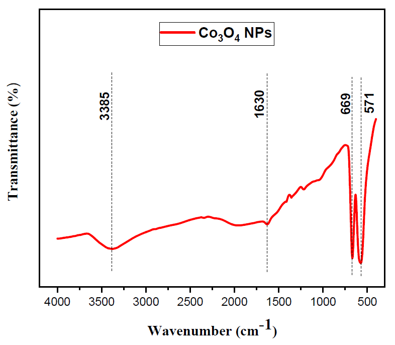
Fourier transform infrared spectroscopy for the obtained product (Co3O4·NPs) was presented in Figure 2. The complete acquired spectra of exhibit two absorption bands at 669 and 571 cm−1, attributed to the (Co–O) vibration modes, as well as a broad band about 3385 cm−1 and a minor band at 1630 cm−1 due to the O–H stretching mode. The sample’s FTIR spectrum reveals two prominent absorption bands at 669 and 571 cm−1, confirming the spinel structure of (Co3O4·NPs). The (Co+2–O) bond’s stretching vibration mode, in which Co2+ (3d7) is located in the tetrahedral hole, is represented by the peak that formed at 669 cm−1. It is possible to attribute the band at 571 cm−1 to the (Co+3–O) bond (where Co+3 (3d6) in the octahedral hole [31,32].
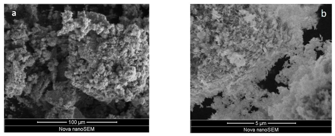
The morphology of the cobalt oxide nanoparticles (Co3O4·NPs) modified by polyethylene glycol (PEG) as surfactant was examined by scanning electron microscope (SEM) (Figure 3). The SEM micrograph at different magnifications shows that the nanoparticles have well uniform spherical shape with a narrow size distribution of 15 nm, which the result of X-ray diffraction (XRD) confirmed by using Debye-Scherrer equation.
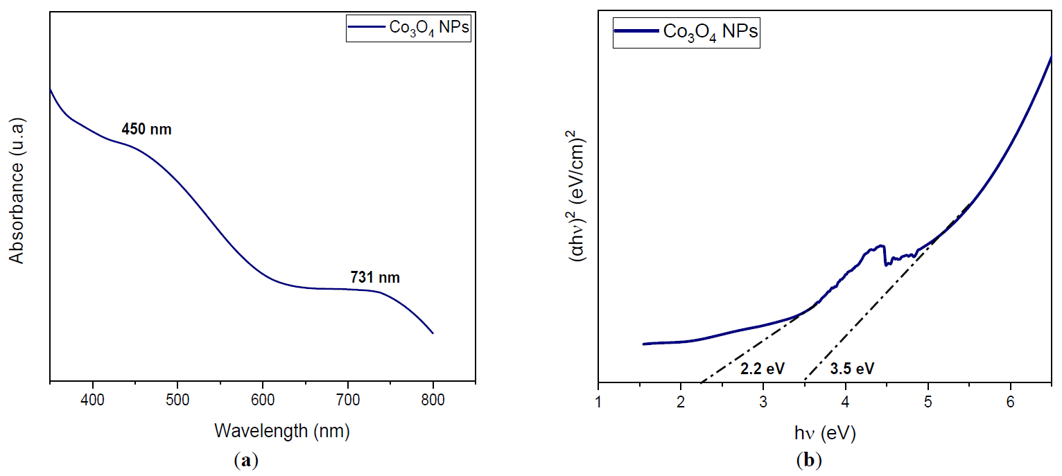
The continuous oscillation of electrons in the conduction band caused by the interaction of an electromagnetic field is the source of light absorption by metallic nanoparticles [33]. Consequently, UV-vis spectroscopy was used to do the optical investigation of the cobalt oxide nanoparticles. UV-vis spectroscopy was used to evaluate the optical characteristics of the as-synthesized Co3O4 nanoparticles, which were created by a sonochemical process using polyethylene glycol (PEG) as a surfactant. The results display a typical absorption spectrum of cobalt oxide nanoparticles (Figure 4). The illustration clearly shows two broad absorption bands. The first absorption band (I) extends from 600 to 800 nm and is associated with the O2− to Co3+ charge transfer process (valence to conduction band excitation). The second absorption band (II) extends from 350 to 500 nm and is associated with the O2− to Co2+ charge transfer process (with the Co3+ level situated beneath the conduction band) [34]. It is noteworthy that absorption is lower in the long wavelength (visible light) region and higher in the short wavelength (blue-violet) region.
The amount of energy needed to excite an electron from the valence band to the conduction band is known as the material’s band gap energy [35]. To estimate this value, the Tauc relation can be applied:
where Eg is the band gap energy, B is a constant, α is the absorption coefficient, n is the factor that depends on the kind of electron transition, h is the Planck constant, and ν is the photon’s frequency. n = 2 for Co3O4 nanoparticles since direct-allowed transition bands are used [36]. From the Tauc plot, two absorption peaks result in two band gap (Eg) values 3.5 and 2.2 eV for the cobalt oxide nanoparticles as prepared using polyethylene glycol as a surfactant.
Additionally, based on this blue shift, it can be deduced that the particles exhibit a decrease in the mean diameter of the quasi-spherical Co3O4 nanoparticles synthesized by using polyethylene glycol (PEG) as a surfactant, which SEM images investigation also verified. Which gives a better energy band gap (Eg) of cobalt oxide nanoparticles (Co3O4·NPs) with the presence of surfactant polyethylene glycol (PEG).
In general, PEG acts as a surfactant for produced cobalt oxide nanoparticles, and when the size of the nanoparticles decreases, the bandgap rises because of quantum size effects. The result could be used for a variety of applications, such as catalysis, energy storage, and sensors.
Analytical grade reagents, cobalt chloride hexahydrate (CoCl2·6H2O), sodium hydroxide (NaOH) and polyethylene glycol (PEG 4000) without any further purification.
Co3O4 nanoparticles were synthesized via the sonochemical method. In a typical experiment, Cobalt chloride hexahydrate (11.89 g) and 5 g of PEG were dissolved in 50 mL of distilled water. Then NaOH (2 M) was added drop by drop into the above solution and sonication for 1 h using an ultrasonic bath. Pink precipitate was filtered and washed with distilled water several times and ethanol. It was then dried under a hot air oven at 60 °C for overnight. Finally, the dried solid was calcined in air at 200 °C for 3 h.
In this study, cobalt oxide nanoparticles (Co3O4·NPs) with an average particle size of 15 nm were synthesized via a facile sonochemical route using polyethylene glycol (PEG) as a surfactant. SEM and XRD measurements showed that the obtained (Co3O4·NPs) are uniform in both morphology and crystallite particle size. And FTIR spectra show two main bands about 669 and 571 cm−1 corresponding to the (Co+2–O) and (Co+3–O) which confirm the spinel structure of Co3O4 and the Co3O4 nanoparticles were found to have optical absorption band gaps of around 2.2 and 3.5 eV.
Due to the presence of PEG around the surface of the product, its particle size was small and the degree of agglomeration is very low. We can use the final product in biomedical, energy storage, gaz sensors and solar energy application due to its small particle size and ferromagnetic behavior.
Finally, the synthesis of cobalt oxide nanoparticles (Co3O4·NPs) by the sonochemical method using polyethylene glycol (PEG) as a surfactant gives the smallest crystallite size compared to other methods and with different types of surfactants or without surfactant.
I thank the Synthesis and Catalysis Laboratory at Ibn Khaldoun University, Tiaret, Algeria, and Khalifa University in the United Arab Emirates for hosting me during my internship.
G.D. conducted all the experiments and wrote the first draft as part of his Ph.D. A.B. supervised G.D. and revised the text, while N.H. collaborated with G.D. on some of the characterizations.
Not applicable.
This research received no external funding.
The authors declare that they have no known competing financial interests or personal relationships that could have appeared to influence the work reported in this paper.